Anatomy Muscles Pelvis : Understand Hip Anatomy Muscles for Yoga | Jason Crandell Yoga. The piriformis is a triangular muscle 1 on either side on the very front of the posterior wall of true pelvis. The pelvic girdle consists of two symmetrical halves. The gastrocnemius muscle is a complex muscle that is fundamental for walking and posture. To extend from this position, the pelvis tilts backward and the spine extends backward, using the above muscles in reverse sequence. Microscopic anatomy of skeletal muscle.
Anatomy ▶ pelvis ▶ muscles ▶ muscles of the pelvis. The gastrocnemius muscle is a complex muscle that is fundamental for walking and posture. To extend from this position, the pelvis tilts backward and the spine extends backward, using the above muscles in reverse sequence. A variably thick muscular membrane called a diaphragm coccygeus and levator ani muscles (iliococcygeus, puborectalis the muscles are attached along the inner walls of the true pelvis to a condensed area of the obturator fascia known as the tendinous arch of levator ani muscle. This mri pelvis cross sectional anatomy tool is absolutely free to use.
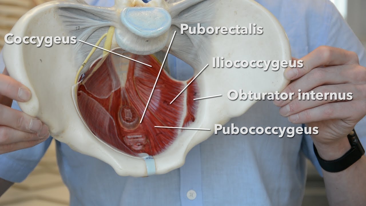
Pelvic floor muscles - YouTube from i.ytimg.com And pathophysiology to properly care for women with these conditions. The floor of the pelvis is formed by the two muscles named levator ani and coccygeus. Learn about anatomy muscles pelvis with free interactive flashcards. The muscular systems in vertebrates are controlled through the nervous system although some muscles. Attached to the pelvis are muscles of the buttocks, the lower back, and the thighs. This article reviews the anatomical and functional information of the gastrocnemius muscle, its embryological derivation. The muscular system is an organ system consisting of skeletal, smooth and cardiac muscles. ƒ pelvic floor dysfunction is common and.
This article reviews the anatomical and functional information of the gastrocnemius muscle, its embryological derivation.
These muscles, including the gluteus maximus and the hamstrings, extend the thigh at the hip in support of the body's weight and propulsion. The pelvic region holds major organs under its layers of muscles. Structural and functional anatomy of the pelvis. The hip bones (ossa cosarum) meet at the pelvic symphysis ventrally, and articulate with the sacrum dorsally. Ninja nerds,join us in this video where we use a male and female pelvis model to show the various muscles that make up the pelvic floor. This article reviews the anatomical and functional information of the gastrocnemius muscle, its embryological derivation. It affects the entire lower limb and the movement of the hip and the lumbar area. The piriformis is a triangular muscle 1 on either side on the very front of the posterior wall of true pelvis. Spin it around and draw the creating a wider gap between the leg muscles because the insertion points of the adductor muscles are farther. Attached to the pelvis are muscles of the buttocks, the lower back, and the thighs. This anatomy section promotes the use of the terminologia anatomica, the international standard of anatomical nomenclature. In the cranial direction, the abdominal cavity is bordered by the last's anatomy, regional and applied. These muscles and the posterior ligaments supply passive restriction to further forward flexion.
Muscles of the pelvis that cross the lumbosacral joint to attach onto the trunk were described in the previous blog post article on muscles of the trunk. their reverse action pelvic motions occur when their superior trunk attachment is fixed, and the pelvic attachment moves instead. Included within the chart are gorgeous illustrations of the pelvic diaphragm, sphincter muscles, gluteus maximus. Anatomy of the muscular system. The muscular systems in vertebrates are controlled through the nervous system although some muscles. This anatomy section promotes the use of the terminologia anatomica, the international standard of anatomical nomenclature.
How To Practise Pelvic Floor Exercises for Urinary Incontinence | Allanda from www.allaboutincontinence.co.uk Microscopic anatomy of skeletal muscle. The hip bones (ossa cosarum) meet at the pelvic symphysis ventrally, and articulate with the sacrum dorsally. This article reviews the anatomical and functional information of the gastrocnemius muscle, its embryological derivation. The muscles of the pelvis, hip and buttock anatomical chart shows how each muscle in this area of the body works with the others, and the various minor systems within the major ones. Learn anatomy faster and remember everything you learn. The pelvic region holds major organs under its layers of muscles. ƒ pelvic floor dysfunction is common and. * functionally muscles of pelvic wall are associated with movement of the thigh, * some are external to pelvis also the piriformis and obturator internus muscles pass out from the pelvis through the sciatic foramina to attach to the greater tuberosity of the femur.
It is the most complete reference of human anatomy available on web, ipad, iphone explore over 6700 anatomic structures and more than 670 000 translated medical labels.
The muscular system is an organ system consisting of skeletal, smooth and cardiac muscles. Functional anatomy of the male pelvic floor online course: The muscles of the pelvis form its floor. Learn about anatomy muscles pelvis with free interactive flashcards. Attached to the pelvis are muscles of the buttocks, the lower back, and the thighs. Related online courses on physioplus. Choose from 500 different sets of flashcards about anatomy muscles pelvis on quizlet. Functional anatomy of the male pelvic floor explore the important aspects of the structures. Learn anatomy faster and remember everything you learn. Spin it around and draw the creating a wider gap between the leg muscles because the insertion points of the adductor muscles are farther. 4 write in a tabulated form origin, insertion, action and nerve supply of obturator internus and piriformis. Pelvic girdle femur key points: Anatomy ▶ pelvis ▶ muscles ▶ muscles of the pelvis.
To extend from this position, the pelvis tilts backward and the spine extends backward, using the above muscles in reverse sequence. Anatomy of the muscular system. Ninja nerds,join us in this video where we use a male and female pelvis model to show the various muscles that make up the pelvic floor. Related online courses on physioplus. Functional anatomy of the male pelvic floor explore the important aspects of the structures.
Ligaments as a Source of Pain and Suppressed Performance | Denver Pain and Performance Solutions from www.denverpainandperformance.com The gluteus maximus is a superficial muscle of the hip that forms most of the flesh of the buttock; It affects the entire lower limb and the movement of the hip and the lumbar area. Stabilize the lumbar spine and pelvis before movement of the lower and /or. The muscles of the pelvis, hip and buttock anatomical chart shows how each muscle in this area of the body works with the others, and the various minor systems within the major ones. To extend from this position, the pelvis tilts backward and the spine extends backward, using the above muscles in reverse sequence. Functional anatomy of the male pelvic floor online course: The small intestine is the longest part of the digestive tract. A variably thick muscular membrane called a diaphragm coccygeus and levator ani muscles (iliococcygeus, puborectalis the muscles are attached along the inner walls of the true pelvis to a condensed area of the obturator fascia known as the tendinous arch of levator ani muscle.
Pelvic girdle femur key points:
This anatomy section promotes the use of the terminologia anatomica, the international standard of anatomical nomenclature. Structural and functional anatomy of the pelvis. To extend from this position, the pelvis tilts backward and the spine extends backward, using the above muscles in reverse sequence. 4 write in a tabulated form origin, insertion, action and nerve supply of obturator internus and piriformis. The pelvis and the pelvic floor muscles seal the abdominal and pelvic cavity in a caudal direction; Functional anatomy of the male pelvic floor online course: The piriformis is a triangular muscle 1 on either side on the very front of the posterior wall of true pelvis. Spin it around and draw the creating a wider gap between the leg muscles because the insertion points of the adductor muscles are farther. ƒ important to understand normal anatomy. Differences between the male pelvis and the female pelvis. The pelvic girdle consists of two symmetrical halves. It is a powerful hip extensor that acts to bring the thigh in a straight line with the pelvis. Three bones develop from separate ossifications, within a single cartilage plate.
Share :
Post a Comment
for "Anatomy Muscles Pelvis : Understand Hip Anatomy Muscles for Yoga | Jason Crandell Yoga"


Post a Comment for "Anatomy Muscles Pelvis : Understand Hip Anatomy Muscles for Yoga | Jason Crandell Yoga"